Physiology of Visual Perception pdf (vision perception)
Today we will be talking about the Physiology of Visual Perception pdf (vision perception), now you might wonder in the course on perception why am i limiting this to visual perception but visual perception per se is the most important or probably it is better to say the most investigated form of perception obviously you have all the senses you have the site you have heard you have an old faction that smells you have gustatory that is the taste you have tactile that is related to touch, All of these senses basically contribute to this perception but the most investigated area or something that has been more studied in cognitive psychology is this area of visual perception the eyes or say for example the most important information that probably we get out of our surroundings is through the eyes and in that sense that kind of skewness in the amount of research that is done in each of these areas.
Cognitive Science has updated the old adage that, “Eyes are the windows to the soul” to Eyes are the windows to the brain. Visual perception is one of the dominating fields of research in cognitive science. Welcome to the world of Visual perception. Indeed we live in a visual world but still, research in visual perception is inadequate to completely explain the process. To start, with this article we will begin with the functionality of photoreceptors (Rods and Cones) in the eyes. We will also talk about how neurochemical impulses are processed by various parts of the eyes, and how these messages are relayed via optic chasm to various areas in the visual cortex that leads to visual perception (color, form, motion, and so on).
Also traditionally in courses in cognitive psychology visual perception is generally something which is given slightly more importance I am not saying that I am following the convention here but we will begin with understanding more details about visual perception in the end and if time permits we may we might talk about auditory perception and related processes as well that said we will talk about the basic part of the first entrance into what visual perception as a process is like so how does this really begin we’ll try and understand the physiology of visual perception function or say for example how do our eyes see what really helps our eyes see and what are the very basic processes I am not going to go into a lot of detail about each of this the idea is just to give you a flavor of what are the constituent processes in visual perception.
Visual perception: The Beginning
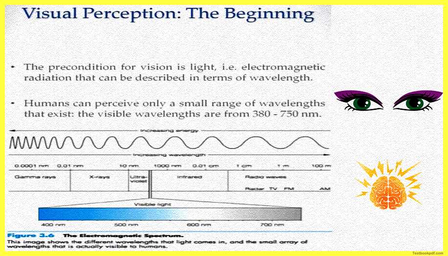
So, let us begin the beginning point or the initiating point of visual perception is basically the registering of light in your eyes so the precondition for the fact that you can see anything for that matter is the presence of light if there is light you see if there is no light you don’t so it is kind of a sort of a binary there
What is light?
Light is basically electromagnetic radiation that can be physically described in terms of wavelength there is this range of wavelength which we are more sensitive to and that basically forms what is called visible light for us.
Humans can perceive only a very limited range of the wavelength of these electromagnetic radiations and this visible wavelength basically falls between 380 to 750 nanometers.
Here you can see this range of visible light so there is this electromagnetic radiation and right from 380 nanometers to 750 nanometers is what we can actually see so any electromagnetic radiation or the electromagnetic radiation in this wavelength is what constitutes our visible spectrum.
How does the process of vision really begin?
How does light contribute to us seeing?
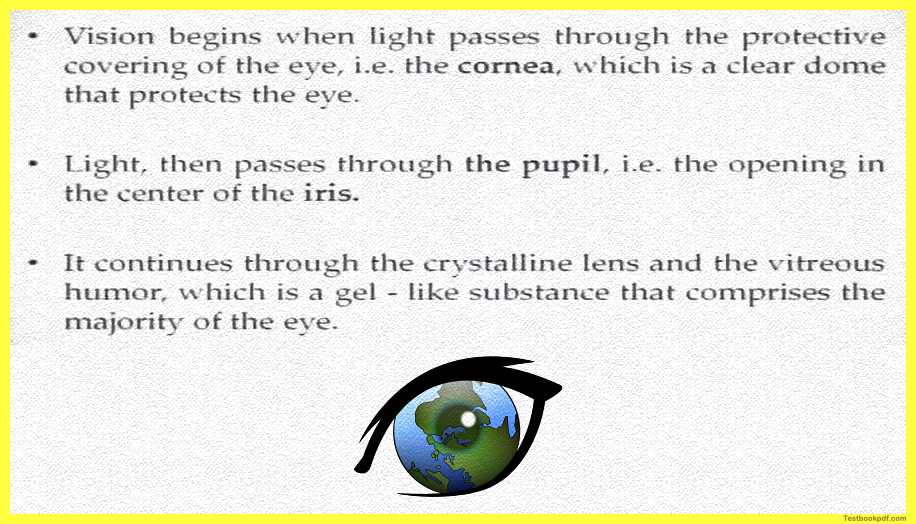
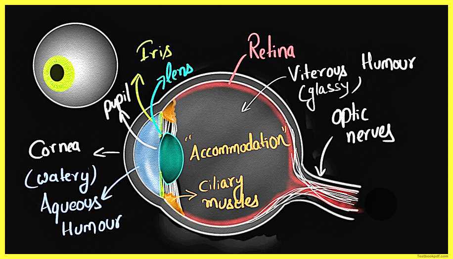
So vision basically or our sensation of anything in this world through the eyes starts when light passes through this protective covering of the eye the top part of the eye which is the cornea which is a clear dome that protects the eye we’ll talk about that I can actually show you the figure and come back here this is what the eye actually looks like.
You can see there are these different parts here you can see that there is a pupil there is cornea there is a part called iris there is the posterior chamber wherein the lens is situated and then there is this a ciliary body which has those muscles, the eye is filled with some substance which is called the vitreous humor at the back you can see there are things like the retina there is the fovea and there is an exiting nerve which is called the optic nerve.
The Human Eye
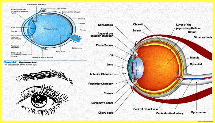
So again I am just taking names of these different areas we will talk about them in much more detail as we go ahead.
What is the cornea?
The cornea is basically a clear dome that is protecting dye something that is the outer layer of the eye then through this cornea the light passes through the pupil, the pupil is basically the opening in the center of the iris you can again refer to the figure here you can see that there is this pupil which is the opening in that thing called the iris you can see that already in the figure this movement.
So, light kind of so it starts through the cornea and passes through the pupil which is basically the opening in the center of the iris and it continues through this crystalline lens which you saw and the vitreous humor which is a gel-like substance which is filled in the eye and then it reaches the back.
So, you see the eye starts at the cornea goes through the pupil passes through the lens passes this entire gel-like vitreous humor, and goes to the back is where the real processing of the light starts so the light then focuses on what is called the retina you might again refer to the figure at the back there is this retina, the retina is basically where the electromagnetic energy.
That is light is transduced that is converted into neural electrochemical impulses if you are interested in what transduction is any signal from the external world you might remember in the psychophysics class we have talked about it any signal from the external world any sensation needs to be converted into a signal that can be understood by the brain so the electromagnetic radiation which is light needs to be converted into neural impulses so that the eyes or the brain, more importantly, can make sense of it.
So the light passes through the cornea goes through the pupil passes the lens and the switches humor goes and falls back at the retina where this is converted into electrical impulses that is where the brain starts making sense of this the vision is most acute at the fovea is a very small thin region of the retina which is basically at the size of a Pinay just like the size of the head of the Pinay.
The retina basically contains layers of neuronal tissue this neuronal tissue basically includes different kinds of cells, cells as the ganglion cells the amacrine cells horizontal cells bipolar cells, and then also photoreceptor cells called the rods and the cones we will.
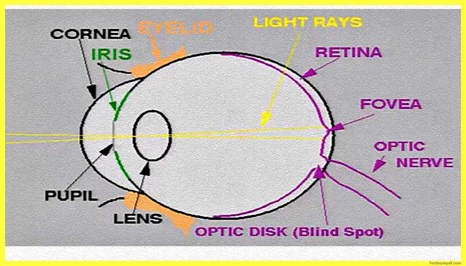
So here you can see a more clear picture of the cornea the iris you see the pupil is there there’s the retina at the back and you’ll see that bulge just like a pin the head of the pin that is what the fovea is.
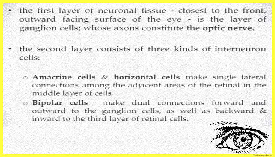
And here there is this layering of these different kinds of cells so the first layer of the neuronal tissue at the retina is closest to the front outwards facing surface of the eye so the first layer is the layer which the light falls first so outward facing surface of the eye this first layer is basically the layer of what is called the ganglion cells, the axons of these ganglion cells if you remember the chapter on the perception the axons are actually the part which kind of protrudes from the cell body the axons of these cells constitute combined together to form what is called the optic nerve.
So this is the outer facing surface of the cell the ganglion cells which form the optic nerve I will show that show the figure to you now so you can see that see there are these ganglion cells in layer number three and the axons of which are forming what is called the optic nerve.
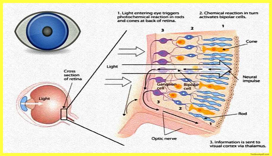
Coming back the second layer consists of three kinds of inter-neuron cells these cells are the anticline and the horizontal cells and the bipolar cells the American cells basically make single lateral connections to adjacent areas of the retina in the middle layer of the cells the bipolar cells make dual connections they make connections forward and outward to the ganglion cells as well as backward and inward to the third layer of these retinal cells.
So you see these connections now you can see the bipolar cells being connected to the inward rods and cones and also to the ganglion cells so they are basically connected to both the inside and the outside part the third layer is basically the layer which consists of what are called photoreceptors as you can guess from the name these are the cells which basically convert light energy into electrical energy that is transmitted by the neurons to the brains so this is where exactly this process of transduction is happening there are two kinds of photoreceptor cells rods and cones each eye consists about 120 million rods and around 8 million cones both of these cells have their own specialties I will talk about them in just a moment.
Now rods and cones simply do not only differ in shape but they also differ in their compositions what they are made of their locations and how they respond to light before we move further it might be helpful to see these cells again so you see that there are three layers the first layer or actually the third the layer number three is basically your ganglion cells the layer number two is your bipolar cells connected both inwards and outwards and the layer number one is basically your photoreceptors you can see the rods and cones in different shapes there.
Okay so you can see how light is really moving light is energy entering the eye and it’s triggering photochemical reactions in rods and cones at the back of the retina chemical reaction, in turn, activates the bipolar cells from the information of the bipolar cell is going to the ganglion and from there it is going to your optic nerve and via the optic nerve, it actually transmits to the brain.
Now, what are these rods and cones doing these rods and cones basically have substances which are called photopigments, photopigments are chemical substances that react to light and transform physical electromagnetic energy which is light into an electrochemical neural impulse that can be understood by the brain now electrical chemical neural impulse we have talked about this in the physiology part where we were talking about neurons action potentials and those kinds of things.
The rods are long and thin photoreceptors more highly concentrated in the periphery of the retina than in the foveal region so rods you will find are more concentrated in the outside regions of the retina and they are basically responsible for night vision and are sensitive to light induction so rods are basically which are more useful when there is less light in night kind of scenarios because they are basically reacting in binary to whether there is light or whether there is darkness or absence of light.
The cones are very short they are slightly thicker photoreceptors and they allow for the perception of colors they are highly concentrated in the foveal region than in the periphery because this is where the light is focused more this is where the acuity of understanding of releasing shapes releasing colors is best so the rods are more sensitive to black and white the colors are more sensitive to different and the cones are more sensitive to different kinds of colors.
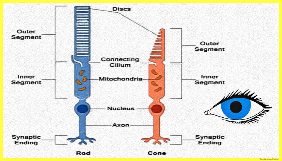
Here you can see the difference basically in rods and cores you see they are not only structurally different and they are also functionally different as we discussed already.
Now the rods cones and the photo pigments could actually not do their job if they were not really connected somehow to the brain so we have to talk about how all of this really connects to the brain as well so the neurochemical messages that are generated in this section of the eye produced by the rods and the cones travel via these bipolar cells to the ganglion cells.
The axons of the ganglion cells in the eyes collectively form what is called the optic nerve for that particular eye you can see that in the figure that we saw earlier so this is how this communication is actually happening.
The optic nerve basically of the two eyes joins at the base of the brain to form something called the optic chiasm basically or at this point what happens is that the ganglion cells from the inward or the nasal part of the retina cross through the optic chiasm and extend to the opposite hemisphere of the brain so.
For example, I can show you show this to you through this figure you can see this figure here.
you will see that there is an inward or nasal part and there is an outward which is basically the retinal part so the inward part basically is where this crossover is happening and so input from one eye is going to another hemisphere input from the say for example from the left eye is going to the right hemisphere input from the right eye is going to the left so this cross over point is called the optic chiasm.
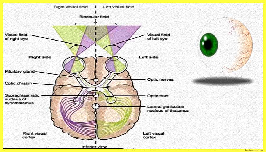
Just going back so at this point at the optic chiasm the ganglion cells from the inward or the nasal part of the retina cross over through the optic may extend to the opposite hemispheres of the brain the ganglion cells from the outward or the temporal area so the inward area is called the nasal area the outward area is called the temporal area which is actually near the temples the temporal area of the retina goes to the hemisphere on the same side of the brain so this is how this crossover happens the lens of each eye basically inverse the image so you might have done elementary physics in your class sixth or seventh you might have known that a particular lens kind of forms an inverted image of the world as it projects this image and the same happens with the lens of the eye so it projects an inverted image to the retina after being routed through the optic chasm about 90 percent of the ganglion cells then go to what is called the LGN or the lateral geniculate nucleus this is a set of nuclei in the thalamus.
So thalamus is the basic region of the brain wherein this is going from the thalamus what happens is that the neurons carry information to the primary visual cortex which is the first area in the occipital cortex that kind of receives this information primary visual cortex in the occipital lobe of the brain and this occipital cortex basically is where the detailed processing of this information coming from these from the retina starts to happen so this visual cortex or your occipital lobe it contains several processing areas each of these areas really carry out different kinds of analysis on the incoming visual informationay and these areas can differ into how they process the intensity of light how they process the quality how they process the color the depth information the location of something the kind of pattern and there is the form.
Visual perception is a really complex activity I can take an example I can say that if I am looking towards you I am actually looking at multiple levels of information I am looking at colors and looking at contours that shape I am looking at whether it is a two-dimensional or a three-dimensional image so I’m looking at depth as well I’m making sense of how far you are from me that is distance so I’m doing and I’m also saying, for example, processing whether you’re moving or not isn’t it.
Details of the Human Eye (Names)
So even though visual perception typically might seem rather simple there are so many processes happening at the same time with anything you’re looking at so if you look out and you are kind of able to figure out that there is a car moving on the road you are actually doing a lot of these analyses over there you have the basic shape of the car you have the colors you have the information that is moving you have you might get to know that what kind of car it is your kind of doing that kind of analysis as well so doing these multiple levels of analysis on whatever visual information is coming to your eyes via light.
let us have a look at this figure this is how this is really arranged you have a binocular field wherein both eyes are getting the input but you also have the right visual field and the left visual field so the right visual field is predominantly basically what the right eye is looking at the left visual field is predominantly what the left eye is looking at.
You can see here in this figure that part of the information from each of the visual fields goes directly to the contralateral hemisphere contralateral is basically the other side of the optic chiasm so you can see in this figure the information in the right visual field goes first to the left visual cortex the information in the left visual field goes first to the right visual cortex i am saying first because generally these contralateral connections of these ganglion cells are supposed to be are found to be faster than the ipsilateral basically the same side connections so this kind of also leads to really interesting or important effects we will talk about them in due course when we are actually talking about more things about visual perception.
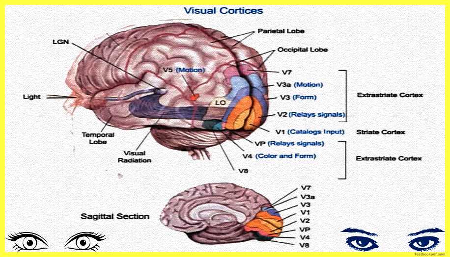
For reference here is the figure which presents what is called the visual cortices.
you can see that there is this lateral geniculate nucleus which is connected to the brain you can see light is entering through the eyes it is kind of through the optic nerve reaching this area called the lateral geniculate nucleus that is what your thalamus is about and from there the information is being related to what is called the occipital lobe which is low which has to do with vision and you can see these so many areas here.
Each of these areas is referred to as so v 1 to v 8 you can see and you can see what these what the specializations of each of these areas say for example i am just reading out for you areas from v1 basically is called this triad cortex it catalogs whatever input is coming in area v2 basically is more responsible for relaying the signal to higher association areas v3 basically processes form v3a processes motion so these are both the areas are in the same place then you have areas like v4 v5 v6 v7 v8 those kinds of things.
Basically so this is how the processing of information starting from light till coming to the brain really happens this is basically all about the physiology that we will be learning in this particular article we will go on to how these different areas in the occipital lobes perform visual processing in the coming article we’ll talk about form perception depth perception constancies those many different things and how basically you make sense of a particular visual stimulus all of that in the coming article on perception thank you.
Read also:
Signal Detection Theory Psychology Pdf
Psychophysics Measuring Sensation (Pdf)
Click here for Complete Psychology Teaching Study Material in Hindi – Lets Learn Squad
