Neurons And Synapse Pdf Free Download
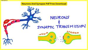
In this article, we will be going to talk about neurons and synaptic transmission, Neurons And Synapse Pdf Free Download and we will discuss the topic with Images So, Let’s start.
What is Neuron?
Neurons are cells that transmit signals between brain cells. Each neuron consists of a cell body (the soma), dendrites, and an axon. Dendrites receive information from other neurons while the axon sends messages to other neurons. A synapse is where two neurons communicate with each other. There are three types of synapses: chemical, electrical, and hormonal.
What is Synapse?
Synapse is the connection between neurons. It is where information is transferred from neuron to neuron. In humans, synapses are located in the brain and spinal cord. There are two types of synapses: chemical synapses and electrical synapses. Chemical synapses transfer signals using neurotransmitters. Electrical synapses use gap junctions to transmit signals.
This Image List is going to start off by covering –
- Neuron structure
- Synaptic cleft,
- Neurotransmitters,
- The concept of synaptic transmission.
Remember how one of the major themes of this course is the relationship between structure and function.
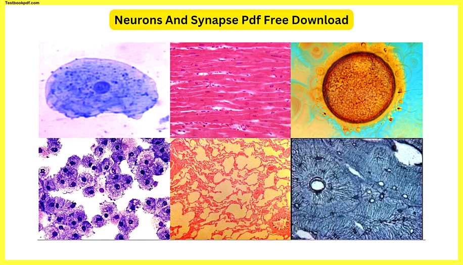
Below you can see all of these different types of tissues that are made up of specialized cells. Most of them have very similar structures inside, but the overall shape of the cell is modified to allow the cell to perform its function. See how many of these types of cells you can identify.
Neurons
Neurons look very different from other cells in the body. Neurons are shaped very similarly to a tree, with many branches and a long trunk. The largest part of a neuron is called the soma o the cell body. Within the soma, we can see the structure called the nucleus which you can easily see in most cells with the aid of a microscope.
If we were to look even more closely inside the body of a neuron, we would notice that it has all the same organelles as a regular cell. It has microtubules, it has ribosomes, it has mitochondria, and all the other organelles that you’re familiar with.
We’re going to look at the slightly bigger picture, though. Neurons function by connecting to other cells and so the more connections they can make, the more effective they’re going to be. These connection sites at the end of the cell that holds the nucleus are called dendrites. We’ll look at the specific role the dendrites play in just a few minutes. What looks like an extra-long dendrite protruding out of the soma is called the axon.
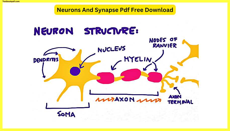
Electrical signals will travel down along the axon away from the soma. Generally speaking, it’s better to have faster conduction of electrical impulses.
In order to speed up the conduction process, we wrap the axon in specialized fatty tissue called myelin. Notice that there are small empty spaces in between the myelin pieces; these spaces are called the Nodes of Ranvier and they play a very important role in the conduction of the impulse along the axon which we’ll talk about in a little bit. At the end of the axon, we have what’s called the axon terminal.
The axon terminal is where this neuron will connect to the next neuron in the pathway. The signal will be passed from the first neuron to the next neuron and then on to whatever neurons are left down the way. However, we have a small problem. If you look at this scene a little bit more closely you’ll notice that the two neurons don’t actually make physical contact with each other. There’s actually a space that’s left in the center like this.
The electrical signal from the first neuron is unable to jump the gap to the second neuron unless other factors come into play. The neuron signal will actually come to a stop right here and this is where our second topic of neurotransmitters and the synaptic cleft are going to come into play. This space in between the two neurons is called the synaptic cleft.
How the signal can jump this Gap?
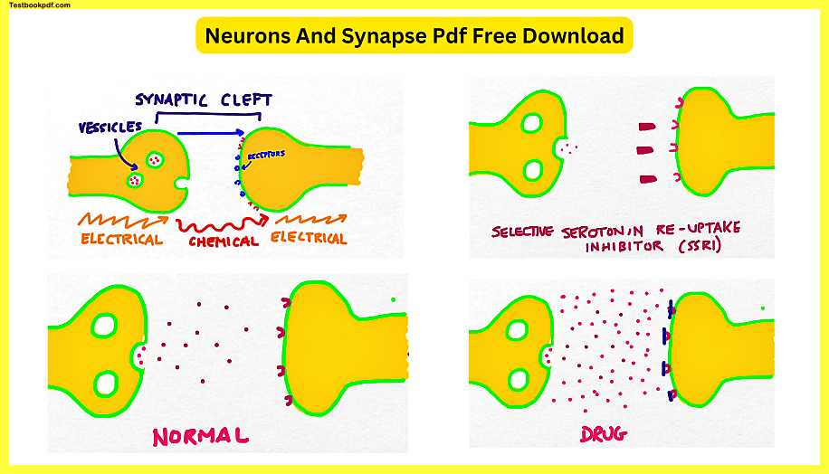
The electrical signal travels down the first neuron and reaches the axon terminal. It then changes forms and becomes a chemical signal. Once the signal has reached the second neuron, it’s then transformed back into an electrical signal again. One of the most interesting topics in all of the neuroscience has to do with what happens during the chemical stage of passing on the signal.
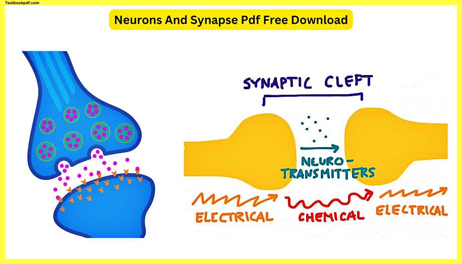
The first neuron releases chemicals called neurotransmitters, which cross the gap to the second neuron and then become an electrical signal again. But, it’s actually a little bit more complicated than this, and so we’re going to look at this a little bit more closely. To understand more about how neuro-transmitters work, we’re going to need to add an additional detail to this drawing.
I’ve now added the cell membrane around each of our two neurons. The neurotransmitter molecules are going to need to somehow pass through the cell membrane in order to get to the next neuron. Inside the first neuron are sealed chambers called vesicles, and inside each vesicle are molecules of the neurotransmitter. The vesicles full of neurotransmitters will be pulled to the edge of the neuron.
Neurotransmitter
Notice that the outside of the vesicle is made of the same material as the outside of the neuron. The vesicle will then dock up against the outer membrane of the end of the neuron in the axon terminal. The vesicle then pops open and deposits the neurotransmitters inside into the synaptic cleft. The neurotransmitter will move in this direction toward the second neuron. We can also call this second neuron the post-synaptic neuron. On the surface of the post-synaptic neuron are a number of small catchment areas which we call receptors.
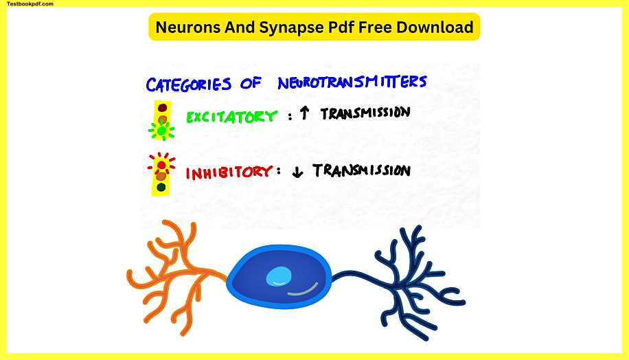
The type of receptor protein on the surface of the post-synaptic neuron will match the type of neurotransmitter that’s been released into the synaptic cleft. Notice that this neurotransmitter will not be able to match up with either of these receptors up here it must dock up against these blue receptors right here. There are two major classes of neurotransmitters; excitatory neurotransmitters and inhibitory neurotransmitters. Excitatory neurotransmitters increase the likelihood of transmission whereas inhibitory neurotransmitters decrease it.
Let’s imagine that this blue neurotransmitter is an excitatory neurotransmitter. The binding of a certain number of molecules of this blue neurotransmitter will prompt the postsynaptic neuron to continue passing along the signal. If it were an inhibitory neurotransmitter, it would prevent the postsynaptic neuron from passing on the signal, or at least decrease the likelihood. The neurotransmitters won’t be bound to the postsynaptic neuron forever, however. Sometimes they’ll be degraded by an enzyme, or sometimes they’ll simply come off on their own. These extra molecules of neurotransmitter are usually reabsorbed into the presynaptic neuron through special channels.
So, in summary, the presynaptic neuron releases a neurotransmitter into the synaptic cleft, which crosses over to the postsynaptic neuron, attaches to receptors, and then prompts the signal to be passed on or inhibited. The signal goes from being electrical to chemical, and then back to electrical again. This is how most neurons communicate with each other. There are other structures called gap junctions that allow neurons to communicate only with electricity, but they’re a little bit rare. The amount of each neurotransmitter present in our synapses at any given time can significantly impact how we feel and how we behave.
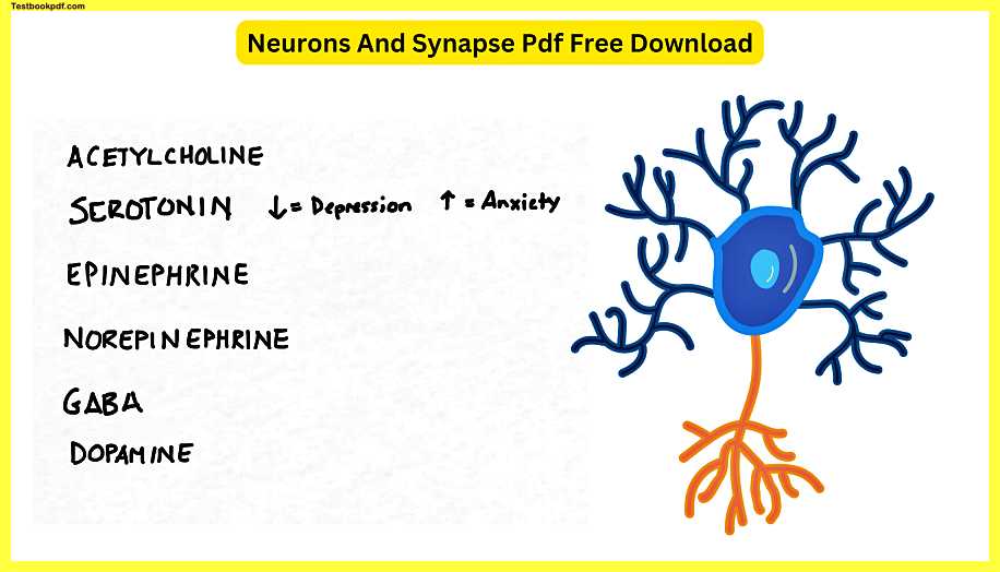
The neurotransmitters that you’ll probably hear about more often are acetylcholine, serotonin, epinephrine, norepinephrine, GABA, and dopamine. Dopamine is going to be one of the neurotransmitters that we’re going to focus on in this class because it is one of the most addictive substances known to man. Depending on whether you have too much or not enough of each of these neurotransmitters and in what combination, it can significantly change a person’s behavior. For instance, low serotonin is associated with depression whereas highly elevated serotonin has been associated with anxiety, specifically social anxiety.
Let’s say for instance that this pink set of dots represents serotonin. The longer the serotonin is present in the synaptic cleft, the more of an effect it will have. In someone who is depressed, the serotonin doesn’t hang around long enough in order to really work. This might occur for a number of different reasons; the serotonin might be degraded by enzymes very quickly, or the person might also make only a little bit of serotonin, to begin with. A common way of treating depression is to employ a selective serotonin reuptake inhibitor also known as an SSRI.
SSRI
The SSRIs block the receptors on the postsynaptic neuron and occupy them thereby allowing the remaining serotonin to spend more time in the synaptic cleft. Common SSRIs in the United States include Zoloft, Paxil, and Prozac. Another neurotransmitter that you hear a lot about is dopamine. Dopamine is also associated with both pleasure and with movement; if you have too little dopamine you often get symptoms like Parkinson’s disease, whereas if you often have too much dopamine you can be at risk for addiction. Many recreational drugs work by manipulating the amount of dopamine in your brain. However, dopamine levels can be manipulated by something as mild as eating a bar of chocolate or playing your favorite game.
Let’s say you get home from classes and all you can think about is how hungry you are and you really want a snack. The minute you start smelling your snack or picking it up in your hand. Dopamine begins pouring out of your neurons into the many synaptic clefts in your brain.
Interestingly enough, the amount of dopamine in your brain is usually at its peak as you ANTICIPATE beginning the enjoyable activity, not when you actually are doing it. If there are more dopamine molecules in your synaptic cleft than there are receptors, the dopamine hangs out for a while before eventually being degraded and reabsorbed. This is what makes many activities feel enjoyable.
Let’s compare a normal amount of dopamine present in the synaptic cleft to that which would be present under the use of illegal drugs such as heroin. Certain drugs cause the body to produce many, many, many more times the normal amount of dopamine. This produces intense effects which can be extremely addictive. Some drugs will take the next step of blocking the receptors on your postsynaptic neurons thereby not only increasing the amount of dopamine that is present but preventing it from being effectively removed.
